No products in the cart.
Advanced Myofascial Techniques: Speaking to Mechanoreceptors
by Bethany Ward & Larry Koliha
Mechanoreceptors are turning out to play an important role in low back pain. The better we understand these sensors, the more specifically we can create meaningful interventions for clients. This article discusses two common mechanoreceptors — Interstitial and Ruffini Receptors — and demonstrates how you can work with them to affect chronic pain and dysfunctional muscle patterns. The techniques presented are from Advanced-Trainings.com’s Advanced Myofascial Techniques series of workshops.
The Interstitial Receptors
Although they also respond to mechanical pressure, interstitial receptors are more commonly known for their nociceptive, or pain-sensing qualities. Studies suggest that these pain receptors can get bad habits. In the presence of extended pain and certain neuropeptides, they can become overly sensitive, producing stronger and more chronic firing than is commensurate with the stimulation.
If hypersensitive interstitial receptors are communicating inappropriate pain messages, can firing levels be reset? It appears that myofascial interventions may be able to “speak” to these intrafascial mechanoreceptors, which are closely linked to the autonomic nervous system (ANS). Research suggests that manual stimulation can alter proprioceptive input to the central nervous system, changing the tonus regulation of the tissue (Schleip, 2003).
Interstitial Receptors: Frozen Hose Technique
To recalibrate interstitial receptors to reestablish more functional firing patterns, go where the mechanoreceptors are most abundant — the periosteum, intermuscular septa, interosseous membranes, or other fascias connected to bone. There seem to be an equal number of interstitial receptors that respond to low threshold pressure as to high pressure, so both rapidly changing and sustained pressure may be effective. This said, engaging the ANS may have a coherent effect, so we recommend taking a strong, slow, sensitive approach, working deeply with the periosteum while looking for signs of manageable ANS activity.
The following technique works with the periosteum of the femur via the iliotibial band. The iliotibial band makes a good handle for this work because “for much of its length, the iliotibial tract is anchored to the femur by the lateral intermuscular septum” (Hollinshead, 1997). A cross-section of the thigh (Image 1) shows how the iliotibial band connects not only with the surface structures of the fascia lata, but also goes deep between the vastus lateralis and biceps femoris, wrapping around the periosteum of the femur.

Image 1: This thigh cross-section shows how the fascia of the illotibial band is continuous with the periosteum of the femur, via the septum between the vastus lateralis and biceps femoris.
The Technique
In the Frozen Hose Technique, the client is sidelying with shoulders and hips stacked vertically and knees bent. Wrap your hands around the iliotibial band, with finger pads gently sinking in at the edges (Images 2 & 3). With a firm (but not pinching) grasp, slowly roll the iliotibial band anteriorly and posteriorly. If it doesn’t want to roll in one direction, attempt to free it by rolling it that way and waiting for a release. Once it is less restricted, roll the iliotibial band again but take it in a direction that creates a stretch away from the bone below. Wait for a release in the tissues. Although your grasp is firm, this technique should not be painful. Use a sensing touch and check with your client to make sure the intervention is well within their comfort level.
This technique is an effective way to address even the very tightest iliotibial bands. For many clients this area is so tight that simply rolling the tissue away from the bone provides a deep stretch into the periosteum. If there is enough slack, lift the iliotibial band away from the femur as you roll the tissue.
You can also experiment with “crimping” the iliotibial band, by taking your hands in opposing directions (much as if you were trying to break up ice in a “frozen hose” — hence the name of this technique). Keep stretching the iliotibial band away from the bone.
Your intention is to provide a deep, steady stimulation that engages the autonomic nervous system at a noticeable but manageable level. Look for signs of client’s heightened proprioceptive presence, such as slower, deeper breathing. It is important that your input is not painful or invasive, overloading the ANS. If you see signs of withdrawal or discomfort, you are not interacting with the tissue in a meaningful way. Your intention is to provide a stimulus that is strong enough to interrupt the habitual response pattern of the interstitial receptors, without reinforcing the current dysfunctional firing patterns.
Although many myofascial therapists have been taught to lean a fist or an elbow into the iliotibial band to lengthen the tissue distally from the hip, this approach may not be producing the expected results. One study calculated that the force needed to produce deformation of high-density fascia (such as fascia lata) was too strong to be achieved by manual manipulation (Chaudhry et al, 2009). Techniques like the Frozen Hose approach may be more effective because they address interstitial receptors, and may affect long-term changes in tissue tone.

Images 2 and 3: In the Frozen Hose Technique, gently sink your finger pads around the anterior and posterior edges of the iliotibial band. Firmly roll the iliotibial band to create a deep stretch in the periosteum below. Wait for a release. If there is enough slack, lift the iliotibial band away from the femur, or create a “crimp in the hose” by taking your hands in oppposing (anterior/posterior) directions. Images courtesy ActionPotential, Inc.
Ruffini Receptors
The Ruffini receptors register mechanical deformation within tissues that regularly experience stretch. Ruffini endings are found in all types of dense connective tissue (muscle fascia, tendons, ligaments, aponeuroses, and joint capsules), but are particularly abundant in peripheral joints, outer capsular layers, and the dura mater.
Similar to the interstitial receptors, addressing the Ruffini organs can lower sympathetic tissue tone via the autonomic nervous system. These sensors are most responsive to techniques that introduce slow, deep pressure (Yahia et al, 1992) and a tangential (or oblique) stretch. Once you engage the tissue with your knuckles, forearm or hands allow your weight to sink in at an angle that creates a shearing of the tissue layer on the layer below. Then wait for the tissue to change under your touch. Go slowly. In addition to changes in tissue quality, look for global changes in the client’s ANS (easier, more expansive breath, or perhaps a relaxing of hands or feet).
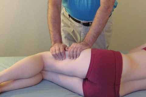
Ruffini Receptors: Seated Back Work with Ball Technique

Image 4: In the Seated Back Work with Ball Technique, engage the tissue on either side of the spine. Allow your weight to sink in at an angle that takes the tissue inferiorly and slides the tissue layer on the one below it. Your pressure should have a slow, melting quality. Image courtesy6 Action Potential, Inc.
The Seated Back Work with Ball Technique is an excellent approach for changing tissue tone of the thoracolumbar fascia, which is so often compressed and tight from dysfunctional postural patterns. Position the client so he or she is seated with hips slightly higher than knees, and knees slightly posterior to ankles. Have the client bend over a medium-sized physioball that is underinflated and has a little give (Image 4). This position decompresses the lumbars while providing support for the low back. Further protect the lumbars by making sure they are anterior to the sacroiliac joints. Lastly, make sure the client’s head is turned to the side, with neck and shoulders relaxed.
Once client is set up, adjust your own stance placing one foot in front of and the other behind your body. Contact each side of the client’s spine with the dorsal side of each hand. Adjust your angle to create a direction that imparts a tangential force in the tissue. Gently sink into the tissue until you feel resistance and slide the lumbar fascia toward sacrum. Your pressure should have a slow, melting quality and should be directed inferiorly, rather than anteriorly. Hook into the tissue layer and only slide at the rate that the tissue allows. Look for systemic changes in breathing, temperature, or relaxation. The client may be able to facilitate this differentiation of layers by gently pushing into the legs and feet, allowing her spine to lengthen superiorly while you coax the outer tissue layer inferiorly.
Conclusion
Our greater understanding of mechanoreceptors is just one way fascia research is growing the field of manual therapy. Additional considerations include how areas of tension change the fascial matrix, the effect of force loading, and the importance of working with fascia as a system. Bringing a variety of creative fascial techniques to your sessions keeps your work fresh and introduces greater possibility for meaningful changes in your clients.
Bethany Ward and Larry Koliha are Lead Instructors for Advanced-Trainings.com, which offers continuing education seminars internationally. They are teaching Advanced Myofascial Techniques workshops for the Head & Neck and TMJ on the Gold Coast, November 8-10.
For more information or to register, go to http://www.amt.org.au/downloads/workshop-registrations/Advanced-Trainings-2014-workshops.pdf
or phone 02 9211 2441
Ward and Koliha also teach at the Rolf Institute® of Structural Integration, Boulder, Colorado and recently presented at the Association of Massage Therapy’s 2014 Conference in Melbourne.
References
Chaudhry H, Schleip R, Ji Z, Bukiet B, Maney M, Findley T. 2008. Three-Dimensional Mathematical Model for Deformation of Human Fasciae in Manual Therapy, Journal of American Osteopathic Association, Vol 108: 379-390.
McMinn R.M.H. 2005. Last’s Anatomy: Regional and Applied, Ninth Edition, Churchill Livingstone.
Rosse C, Gaddum-Rosse P, Hollinshead WH. 1997. Hollinshead’s textbook of anatomy, Lippincott-Raven Publishers.
Schleip R. 2003: Fascial plasticity – a new neurobiological explanation. Journal of Bodywork and Movement Therapies 7(1):11-19 and 7(2):104-116.
Yahia L, Rhalmi S, Newman N, Isler M. 1992. Sensory innervation of human thoracolumbar fascia. Acta Orthop Scand 63(2):195-197.